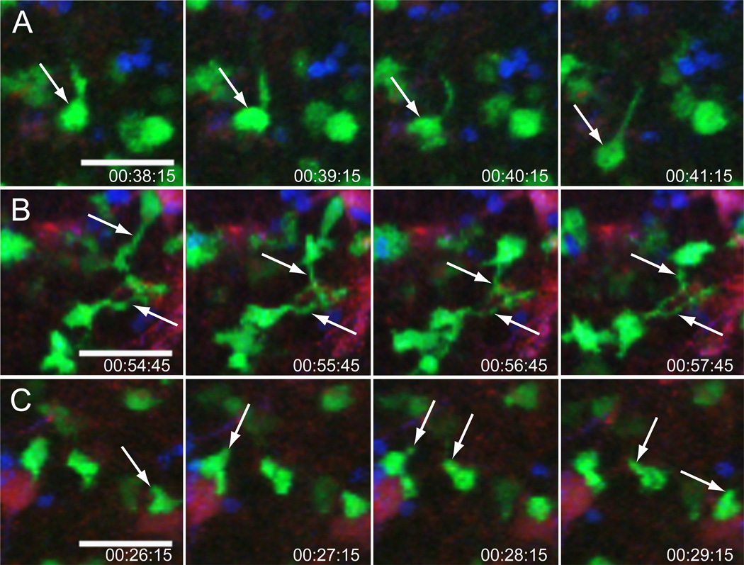
Figure 2. Morphology of GC and examples of GC B cells as imaged by intravital microscopy.
The images shown here are representative of those obtained by intravital time resolved imaging of a popliteal lymph node 9 days after immunization. GC B cells (green), naïve B cells (blue) and FDC cell bodies and dendrites (red). (A) An example of a polarized GC B cell moving directionally (white arrow). (B) Tethered cells with long, anchored cytoplasmic extensions in close association with FDCs. (C) Irregularly shaped GC B cells probing their environment with filopodia. Hapten specific B cells expressing GFP under the direction of the chicken-b-actin promoter were transferred into GFP tolerant recipients. The next day, recipients were immunized s.c. with NP-CGG. Two days before imaging, recipients were injected in the footpad with anti-CD35 Fab fragments conjugated to AL633 to highlight FDCs. They also received wild type naïve B cells that were labeled with Hoechst 33342. Scale bar, 30 µm. Originally published in Hauser et al. Immunity 26:655–667 (2007) and reprinted with permission from Elsevier.

