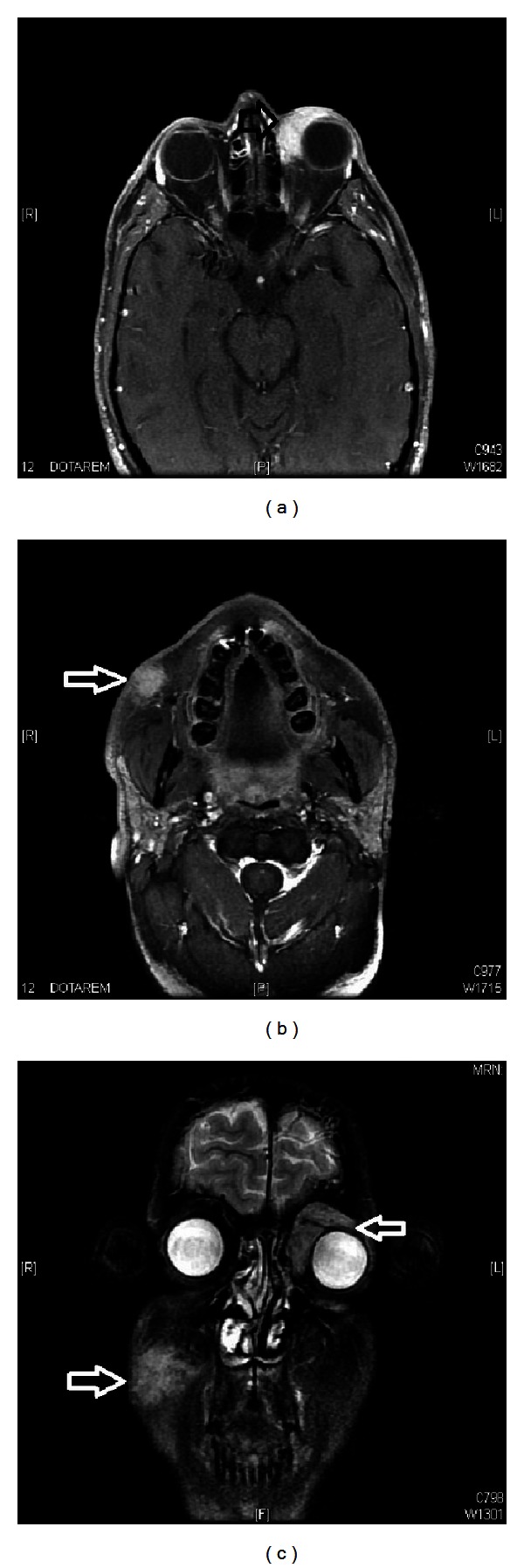
Figure 4.

Magnetic resonance imaging (MRI) ((a) and (b)) axial and (c) coronal images of head showing recurrent mass tissue lesion involving the left eyelid, mainly superiorly and medially progressing deeply to surround the superior and medial recti muscles as well as superior oblique muscle mainly at their insertion on the ocular globe and another mass tissue lesion involving the right cheek included within the subcutaneous tissue showing intense enhancement measuring about 3 × 2 cm without obvious deep extension.
