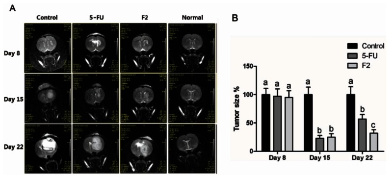
Fig. 2. Human glioblastoma cells (U373MG) were implanted into the right forebrain (n=6/group). (A) Since day 8 after implantation, drugs are intravenously injected every two days at dosages of 35 and 15 mg/kg weight of ginsenoside F2 (F2) and 5-fluorouracil (FU), respectively. T1 weighted magnetic resonance imaging were carried out at 8, 15 and 22 days after implantation of U373MG cells. (B) Tumor sizes were demonstrated as a pixel number by Image J and represented relatively to control. p<0.05, significantly different, compared with control by one-way ANOVA followed by Turkey’s test. The different letters in each day mean difference between groups.