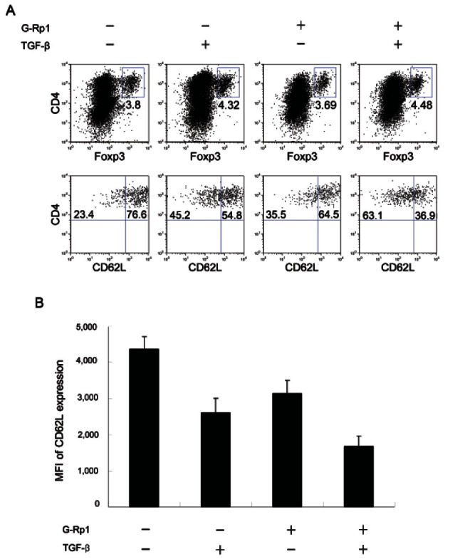
Fig. 3. Expression of CD62L on Tregs stimulated with TGF-β and/or ginsenoside Rp1 (G-Rp1). Activated Splenocytes on anti-CD3 antibody-coated plates were cultured with or without 2 ng/mL TGF-β for 3 d and then 20 μM G-Rp1 for 2 d. (A) Flow cytometry analysis of culture cells costained with CD4 and Foxp3. Gated CD4+Foxp3+ cells were then stained for CD62L. Numbers in quadrants indicate percentage of CD62Llow cells and CD62high cells. (B) Mean fluorescence intensity (MFI) of CD62L expression in regulatory T cells. The results are average of three separate experiments.

