. 2013 Apr;37(2):167–175. doi: 10.5142/jgr.2013.37.167

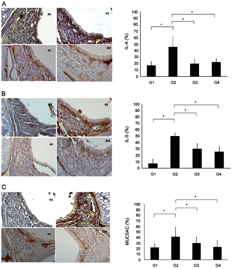
Fig. 5. Immunohistochemical staining (×400). IL-4-, IL-5- and MUC5AC-positive cells were found in the epithelium and submucosal gland, and the numbers of these cells were increased in group 2 (G2). (A) The number of IL-4-positive cells was significantly higher in G2 than in group 1 (G1). Additionally, the number of these cells was significantly lower in group 3 (G3) and group 4 (G4) than in G2. (B) The number of IL-5-positive cells was significantly fewer IL-5-positive cells appeared in G3 and G4 compared to G2. (C) MUC5AC staining revealed a significantly greater number of positive cells in G2 than in G1. The number of positive cells was significantly lower in G3 and G4 than in G2 (*p<0.05). G1, normal saline; G2, ovalbumin (OVA)+saline; G3, OVA+ Korean red ginseng; G4, OVA+dexamethasone (graph: average±standard deviation).

Fig. 5.