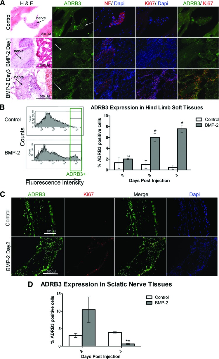
Figure 2.
Activation of ADRB3+ cells associated with the peripheral nerve perineurium after exposure to BMP2. (A): Representative photomicrographs of ADRB3 expression within the soft tissues of the hind limb showed ADRB3+ cells undergoing replication in the perineurial region of the peripheral nerves 3 days after delivery of AdBMP2 transduced cells. Shown are ADRB3 (green), neurofilament (red), and Ki67 (red) in the rightmost column. Arrows indicate the nerves. (B): Flow cytometric analysis of ADRB3 expression within the soft tissues of the hind limb revealed a significant increase in the number of ADRB3+ cells after 3 and 4 days of exposure to BMP2. A representative histogram of the percentage of ADRB3+ cells on day 4 is shown. (C): Representative photomicrographs of ADRB3 expression within the sciatic nerve tissues showed ADRB3+ cells associated with the perineurium and coexpressing the proliferation marker Ki67, 2 days after delivery of AdBMP2 transduced cells. Tissues were counterstained with Dapi (blue). Shown are ADRB3 (green) and Ki67 (red). (D): Flow cytometric analysis of ADRB3 expression within the sciatic nerve tissues revealed a marked increase in the number of ADRB3+ cells after 2 days of exposure to BMP2, followed by a significant decrease after 4 days. For each experiment, six hind limb or nerve samples for each condition were pooled for flow cytometric analysis. Bar graphs show the average of three independent experiments ± SEM. *, p < .05; **, p < .005. Abbreviations: ADRB3, beta-3 adrenergic receptor; BMP-2, bone morphogenetic protein 2; Dapi, 4′,6-diamidino-2-phenylindole; H&E, hematoxylin and eosin; NF, neurofilament; ns, not significant.

