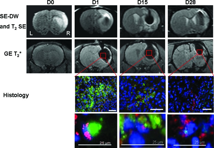
Figure 4.
Labeled human mesenchymal stem cell (hMSC) detection by in vivo magnetic resonance imaging (MRI) and histology. Immediately after middle cerebral artery occlusion (D0), SE-DW images (first row) revealed the right corticostriatal ischemic lesion without any hemorrhage on gradient echo T2*-weighted image (second row). After hMSC intracerebral grafting, at D1, D15, and D28, labeled cells were identified in vivo into both sites of injection (right damaged striatum and cortex) using SE-DW and T2* imaging. A magnetic susceptibility artifact is seen as a signal void (dark area) on T2* images caused by iron particles. For fluorescence microscopy, all of the cell nuclei were counterstained blue with Hoechst. Micrometer-sized superparamagnetic iron oxide (M-SPIO) green fluorescence is directly visualized and merged with a human-specific antibody to nuclear antigen (MAB1281) to identify human cells derived from hMSCs (in red). High-magnification images (bottom two rows) show the colocalization of this staining, corresponding to the presence of viable human cells with intracellular M-SPIO labeling at every time. M-SPIO concentration in the graft sites decreased for 28 days but remained notably detectable by MRI at D28. Scale bars = 50 μm (or 25 μm as noted). Abbreviations: D, day; DW, diffusion-weighted; GE, gradient echo; L, left; R, right; SE, spin echo.

