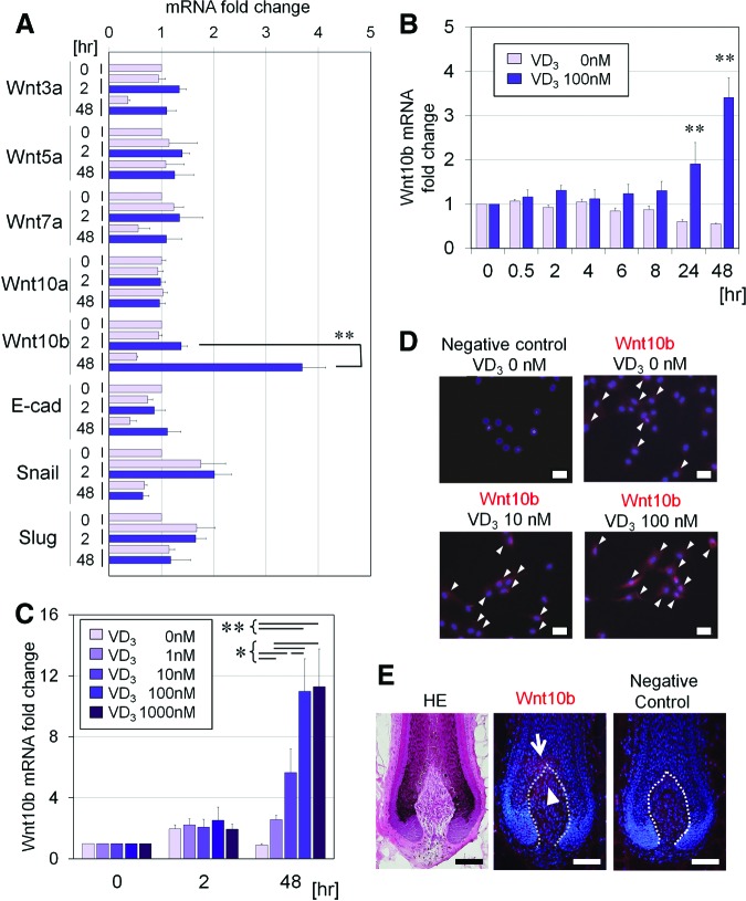
Figure 2.
Effect of VD3 on expression of Wnt-related genes in cultured human dermal papilla cells (hDPCs). (A): Gene expression of Wnt3a, Wnt5a, Wnt7a, Wnt10a, Wnt10b, E-cad, snail, and slug was examined in hDPCs cultured in Dulbecco's modified Eagle's medium (DMEM)/10% fetal bovine serum (FBS) for 0, 2, or 48 hours, with or without VD3 (100 nM). Data are shown as fold change compared with baseline (n = 4). **, Significant differences between pairs (p < .01). (B): Kinetic analysis (0, 2, 4, 6, 8, 24, or 48 hours) of Wnt10b mRNA relative expression in hDPCs cultured in DMEM/10% FBS, supplemented with or without VD3 (100 nM). Data are shown as fold changes compared with the baseline expression (n = 4). **, Significant differences between conditions (with and without VD3), at each incubation time point (p < .01). (C): Dose-dependent effects (0, 1, 10, 100, or 1,000 nM) of VD3 on Wnt10b mRNA expression, in hDPCs cultured for 0, 2, or 48 hours (n = 4). Significant differences between groups, at each incubation period, are shown as * (p < .05) or ** (p < .01). (D): Representative images of immunocytochemical staining for Wnt10b in hDPCs cultured with various concentrations of VD3 (0, 10, or 100 nM) for 3 days. Goat IgG was used as a negative control. Arrowheads indicate Wnt10b protein expression. Scale bars = 50 μm. (E): Representative serial sections of the intact human scalp hair follicles. Shown are HE staining (left), immunohistochemical staining for Wnt10b (middle), and negative control with goat IgG (right). Wnt10b was expressed in the top portion of human dermal papilla (arrowhead) and the adjacent hair matrix (arrow). Scale bars = 100 μm. Abbreviations: E-cad, E-cadherin; HE, hematoxylin/eosin; VD3, 1α,25-dihydroxyvitamin D3.

