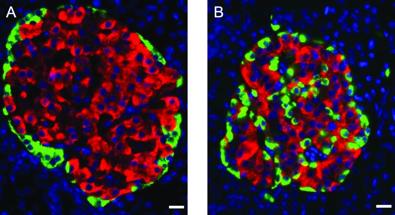
Figure 1.
Comparative organization of endocrine cells in rat and human islets. Pancreatic sections from adult rat (A) and human (B) pancreases were immunostained with antibodies against insulin (red) and glucagon (green). The nuclei were stained with Hoechst 33342 fluorescent stain (blue). Note that in rat islets, glucagon-positive cells surround insulin-positive cells, which is not the case in human islets. Scale bars = 12.5 μm.

