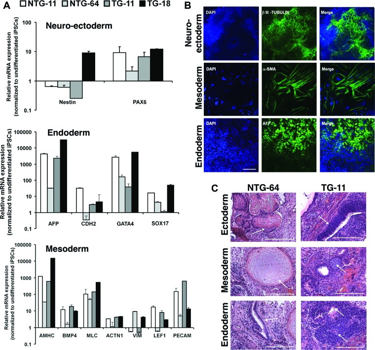
Figure 2.
In vitro embryoid bodies (EBs) mediated differentiation of NTG and TG iPSCs. (A): Differentiated NTG and TG EBs were analyzed by quantitative polymerase chain reaction to detect ectoderm, endoderm, and mesoderm marker expression. Graphs show relative expression to undifferentiated iPSCs in the corresponding sample. (B): iPSCs were stained βIII-tubulin (neuroectoderm), α-SMA (mesoderm), and AFP (endoderm). A fluorescence microscope was used to image the samples, and representative pictures are from a differentiated TG-11 line. Scale bar = 50 μm. (C): Teratoma sections derived from representative NTG-64 and TG-11 iPSCs were collected after 6 weeks, and sections were stained with hematoxylin and eosin. Teratomas contained representative tissues of the three germ layers: pluristratified epithelium (ectoderm), cartilage (mesoderm), and columnar epithelium (endoderm). Scale bars = 50 μm. (A full list of abbreviations is given in the supplemental online data.) Abbreviations: BMP, bone morphogenetic protein; DAPI, 4′,6-diamidino-2-phenylindole; iPSC, induced pluripotent stem cell; NTG, nontransgenic; α-SMA, α-smooth muscle actin; TG, transgenic.

