Figure 3.
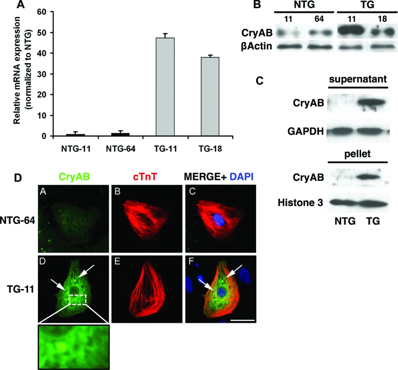
CryAB expression and aggregation in differentiated cardiomyocytes produced in TG EBs. (A): Quantitative polymerase chain reaction analysis of CryAB in NTG-11, NTG-64, TG-11, and TG-18 differentiated induced pluripotent stem cells (iPSCs) compared with undifferentiated iPSCs. (B): Western blots (EB total extract) show CryAB protein expression in EBs derived from NTG and TG iPSCs. (C): Western blots of the detergent-soluble (supernatant) or insoluble (pellet) fractions of EBs show partial accumulation of CryAB protein into the insoluble fraction in TG samples. GAPDH and Histone H3 were used as the loading controls. (D): cTnT immunostaining was used to identify cardiomyocytes in differentiated replated EBs. Only cardiomyocytes derived from TG iPSCs displayed perinuclear protein aggregates (arrows, inset: high-magnification view of perinuclear region). Shown are CryAB (green), cTnT (red), and DAPI (blue) in representative NTG-64 and TG-11. Scale bar = 50 μm. (A full list of abbreviations is given in the supplemental online data.) Abbreviations: CryAB, αB-crystallin; cTnT, cardiac troponin T; DAPI, 4′,6-diamidino-2-phenylindole; NTG, nontransgenic; TG, transgenic.

