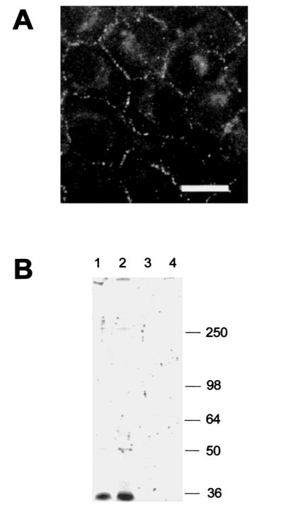
Fig. 4.
Immunofluorescence and phosphorylation analysis of HeLa cells transfected with Cx36. (A) Cx36 immunofluorescence in HeLa-Cx36 cells. Note the strong punctate labeling on contact membranes of the transfectants. There was also weak labeling in the nucleus and the cytoplasm, but no signals were visible in non transfected Hela cells (data not shown). Scale bar: 30 μm. (B). Immunoprecipitation of Cx36 in either Cx36-transfected (lanes 1 and 2) or wild type (lanes 3 and 4) HeLa cells. The beads were incubated with alkaline phosphatase (lanes 2 and 4) or without enzyme after immunoprecipitation. Molecular masses are indicated in kDa at the right.

