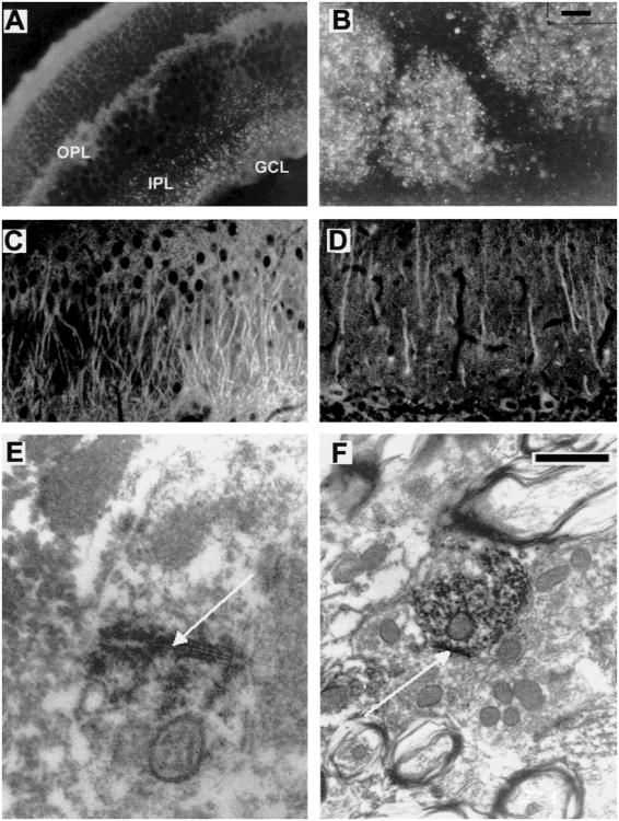
Fig. 5.
Cx36 immunohistochemistry in mouse and rat nervous system at light and electron microscopic levels. (A and B) Cx36 immunofluorescence analysis of ethanol-fixed mouse sections of (A) Retina (punctate staining in the outer plexiform (OPL) and the inner plexiform layers (IPL) ranging to the ganglion cell layer (GCL)) and (B) Bulbus olfactorius (punctate staining in the glomerula). (C and D) Cx36 immunofluorescence analysis of paraformaldehyde-fixed rat sections of (C) Hippocampus, CA3 region: The apical pyramidal cell dendrites in the stratum radiatum exhibit strong immunoreactivity, possibly representing intracellular transport of Cx36 protein. Staining of somata is less prominent; (D) Cerebellum: Immunoreactivity is localized in the Purkinje cells, predominantly in the dendritic processes of the molecular layer. (E and F) Cx36 immuno electron microscopy in the mouse inferior olive. Note the strong punctate labeling at a neuronal gap junction shown (arrow) in E. In F, the Cx36 antibodies labeled the cytoplasm of a dendrite. Scale bar (upper right corner) in (A–D) 30 μm; in E and F: 200 nm.

