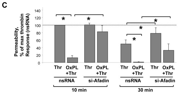
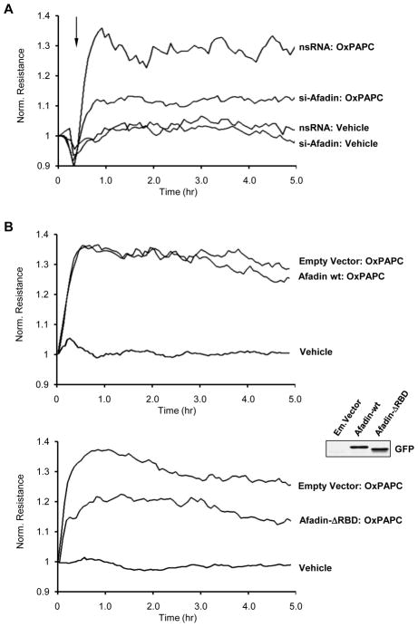
Figure 5. Involvement of afadin in the OxPAPC-induced EC barrier enhancement.

A - Pulmonary EC were transfected with afadin-specific siRNA or non-specific RNA. EC were stimulated with OxPAPC (10 μg/ml) or vehicle at the time indicated by arrow, and TER changes were monitored over 5 hours. Results are representative of five independent experiments. B - EC monolayers were subjected to transfection with full length GFP-tagged afadin (upper panel), or afadin-ΔRBD mutant bearing GFP tag (lower panel). Cells transfected with empty vector served as controls. Inset - expression of recombinant wild type afadin and afadin-ΔRBD mutant in pulmonary EC detected by western blot with GFP antibody. After 48 hrs of transfection, cells were stimulated with OxPAPC. Changes in endothelial permeability were monitored by measurements of TER. Results are representative of four independent experiments. C - HPAEC were transfected with afadin-specific siRNA or non-specific RNA duplexes. EC were pretreated with OxPAPC (10 μg/ml, 30 min) or vehicle prior to thrombin (0.1 U/ml) challenge, and TER was monitored over the time. Permeability increase caused by thrombin stimulation for 10 min (538+/−73 Ohm in comparison to 1164+/−269 Ohm for non-stimulated cells) of EC transfected with non-specific RNA was taken as 100%. Results are representative of six independent experiments.

