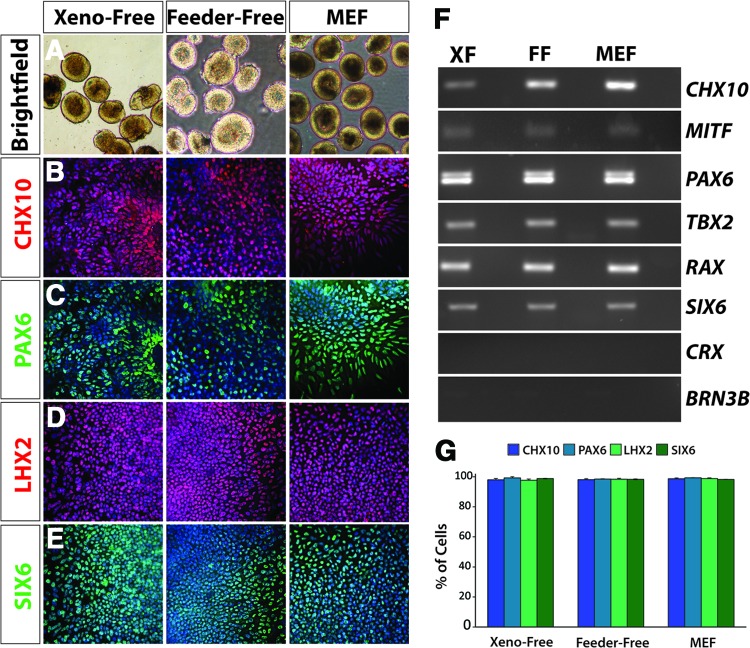
Figure 3.
Derivation of definitive retinal progenitors from human induced pluripotent stem cells (hiPSCs) under xeno-free conditions. (A): Under each of the three growth conditions, retinal progenitor spheres can be isolated under bright-field microscopy using morphological cues. The retinal spheres were identified by a light outer ring around the periphery, a morphological feature absent in nonretinal spheres. Magnification, ×4. (B–E): Retinal progenitor cells expressed characteristic markers such as CHX10, PAX6, LHX2, and SIX6. Magnification, ×20. (F): Reverse transcription-polymerase chain reaction analysis confirmed the expression of these retinal progenitor markers and indicated similar expression levels of cells grown in a nonxenogeneic environment when compared with the cells grown using traditional systems. Markers of more mature cells were not expressed at this stage, including CRX and BRN3, indicating the temporal specificity of these retinal progenitor cells. (G): After manual isolation of retinal neurospheres based on morphological cues, similar levels of expression were observed between samples for each of the indicated transcription factors. Abbreviations: FF, feeder-free; MEF, mouse embryonic fibroblast; XF, xeno-free.

