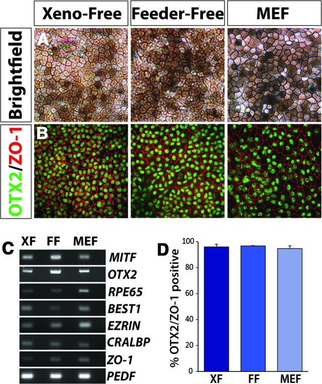
Figure 5.

Retinal pigmented epithelium (RPE) derived from human induced pluripotent stem cells (hiPSCs) under xeno-free and traditional growth conditions. (A): Bright-field microscopy demonstrated the characteristic pigmented, hexagonal morphology associated with RPE specification. This phenotype was first apparent approximately 1 month following the start of differentiation and increased over the next few weeks. Magnification, ×20. (B): hiPSC-derived RPE grown under all three conditions expressed characteristics such as the transcription factor OTX2 and the tight junction protein ZO-1. Magnification, ×20. (C): Reverse transcription-polymerase chain reaction from xeno-free, feeder-free, and MEF cultures similarly expressed a number of RPE-associated genes. (D): Following manual isolation and expansion of RPE derived under each growth condition, comparable percentages of cells coexpressing OTX2 and ZO-1 were observed. Abbreviations: FF, feeder-free; MEF, mouse embryonic fibroblast; XF, xeno-free.
