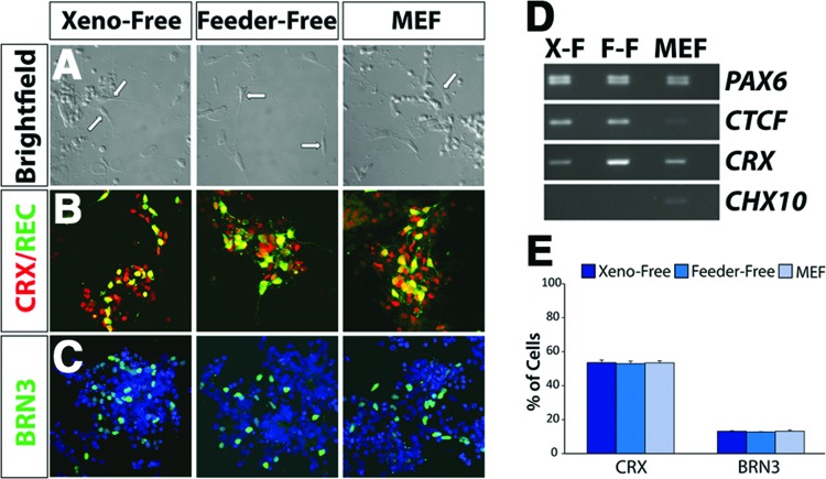
Figure 6.
Neuroretinal cell types derived under xeno-free conditions. (A): After 60 days of differentiation, a subset of cells acquired morphologies of primitive photoreceptor-like cells in vitro, including a unipolar appearance with one short process, as indicated by arrows. Magnification, ×20. (B): At this time point, a subset of cells expressed genes associated with photoreceptor cells, including CRX and RECOVERIN. Magnification, ×40. (C): Other cells expressed markers specific to retinal ganglion cells such as the transcription factor BRN3. Magnification, ×40. (D): Reverse transcription-polymerase chain reaction analysis indicated the expression of genes specific to differentiated retinal subtypes including CRX, whereas the retinal progenitor marker CHX10 is no longer expressed in these cells. (E): Aggregates of cells after 60 days of differentiation exhibited comparable percentages of cells within immunopositive colonies expressing CRX and BRN3, indicative of photoreceptors and retinal ganglion cells, respectively. Abbreviations: F-F, feeder-free; MEF, mouse embryonic fibroblast; X-F, xeno-free.

