
Figure 5.
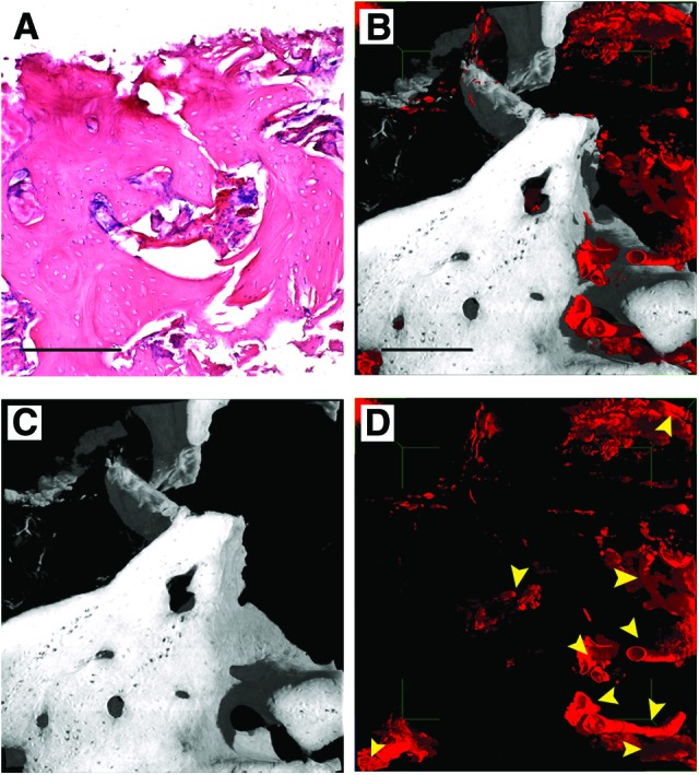
Three-dimensional (3D) images of human mandible control. (A): Histological section with hematoxylin-eosin staining, as a reference. (B–D): Subvolume of the 3D reconstruction from the holotomography investigation. To improve visualizations, in each 3D image all other phases were deleted virtually, except for bone and vessels (B), bone (C), and vessels (D). Yellow arrowheads indicate vessels to distinguish them from possible artifacts. Scale bars = 250 μm.