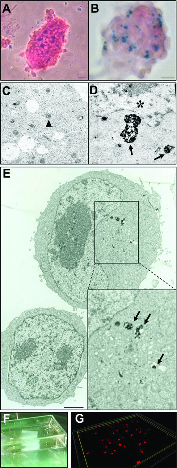
Figure 1.

Evaluation of superparamagnetic iron oxide (SPIO) particle loading on mouse embryonic stem cells (mESCs). Magnetofected- or lipofected-ESC colonies ([A] and [B], respectively) were stained with Prussian blue and contained similar amounts of SPIO. Electron microscopy pictures (C, D) confirmed the presence of iron particles within mESCs using both lipofection ([C, D], 24 hours, 9 μg/ml) or magnetofection ([E], 30 minutes, 7 μg/ml). Triangles, mitochondria; asterisk, nucleus; black arrows, iron-loaded endosomes. Scale bars = 1.2 μm (E) and 400 nm (C, D). (F): Image of a fibrin-based patch (5 × 5 × 1.5 mm), after polymerization of a 200 μl solution into a Lab-Tek multichamber and removal by lid lifting. (G): Example of cell distribution within a section of fibrin gel (300,000 cells per gel) upon 3D reconstruction of confocal images.
