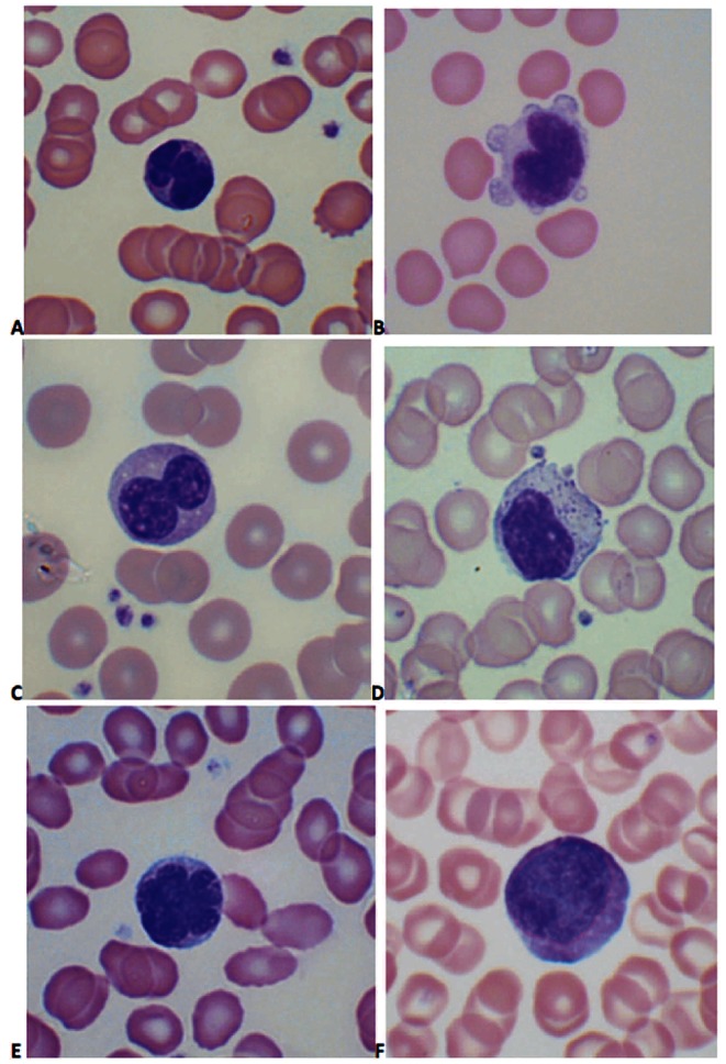
Figure 1.

Lymphocyte morphology in asymptomatic carriers of HTLV-1: (A) convoluted nucleus; (B) cleft nucleus with cytoplasmic blebbing; (C) bi-lobed nucleus; (D) large granular lymphocyte; (E) flower cell; (F) abnormally large cell. (Images were obtained using a Nikon Eclipse E600 microscope, 100x objective, Olympus DP-12 camera; Adobe Photoshop used to adjust contrast and brightness only).
