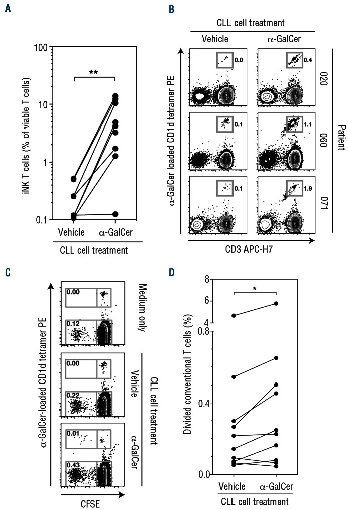
Figure 5.
α-GalCer-treated CLL cells induce allogeneic and autologous iNKT cell proliferation, and enhance conventional T-cell proliferation. (A) Frequency of iNKT cells within healthy donor PBMC following co-culture with allogeneic vehicle-or α-GalCer-treated CLL cells (**P<0.01). (B) Flow cytometry plots showing iNKT cell numbers (percent of viable lymphocytes) after repeated stimulation of patients’ PBMC with autologous vehicle-or α-GalCer-treated CLL cells in the presence of a low-dose of interleukin-7. In a fourth patient, no iNKT cell proliferation was observed. (C) Representative flow cytometry plots showing proliferation of iNKT and conventional T cells following addition of autologous vehicle- or α-GalCer-treated CLL cells, assessed by CFSE dilution. Plots gated on viable CD19CD3+ lymphocytes; numbers represent percent of gated events. (D) Frequency of divided conventional (CD3+,α-GalCer-loaded CD1d tetramer) T cells following stimulation with autologous vehicle- or α-GalCer-pulsed CLL cells (n=10) (*P<0.05).

