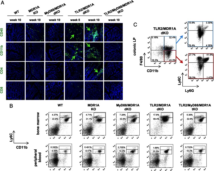
FIGURE 3.
TLR2/MDR1A dKO mice have increased numbers of CD11b+ cells in bone marrow, peripheral blood, and colonic lamina propria. (A) Representative immunofluorescent staining with anti-CD45, anti-CD11b, anti-CD4, and anti-CD8 (FITC, green) of distal colons from 5- or 10-wk-old WT, MDR1A KO, MyD88/MDR1A dKO, TLR2/MDR1A dKO, and TLR2/MyD88/MDR1A tKO mice (n = 3/group), as assessed by confocal laser microscopy (scale bar, 50 μm). Green arrows indicate positive cells. Nuclei were counterstained with DAPI (blue). (B) Flow cytometry of bone marrow and peripheral blood cells isolated from WT, MDR1A KO, MyD88/MDR1A dKO, TLR2/MDR1A dKO, and TLR2/MyD88/MDR1A tKO mice (6 wk old; except WT: 9 wk old; n = 3/group) stained with V450-conjugated anti-Ly6C and streptavidin-coupled FITC detecting biotinylated anti-CD11b. Representative dot plot presents the incidence of CD11b+Ly6C+ cells in the total population. (C) Flow cytometry of colonic lamina propria cells isolated from TLR2/MDR1A dKO (12 wk old; n = 3/group), purified with anti–CD11b-PE magnetic beads, and stained with anti–F4/80-allophycocyanin-eFluor 780, anti–Ly6G-FITC, and anti–Ly6C-V450. Representative dot plot shows the incidence of CD11b+F4/80+ versus CD11b+F4/80− cells in the purified population (left) with further analysis based on surface expression of Ly6C and Ly6G (right). Experiments were repeated at least twice.

