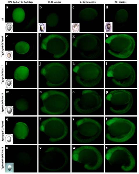
Fig. 2.
Developmental time course of EGFP expression in transgenic embryos. AB embryos were collected at the same time points from either 50% epiboly through the 30+ somite stage as the transgenic lines. EGFP fluorescence was not detected in any of the AB embryos (a–d). EGFP expression was present from the bud stage through the 30+ somite stage in the Tg(h2afv:EGFP)nt13 (e–h), Tg(tbp:EGFP)nt17 (i–l), and Tg(eef1g:EGFP)nt15 lines (q–t). EGFP expression was detected in the Tg(tbnl:EGFP)nt16 and Tg(bactin1:EGFP)nt14 lines from the 10–14 somite through the 30+ somite stages (Panels m–p and u–x, respectively) Embryos that are difficult to see in the fluorescent field have an inset of a bright field image

