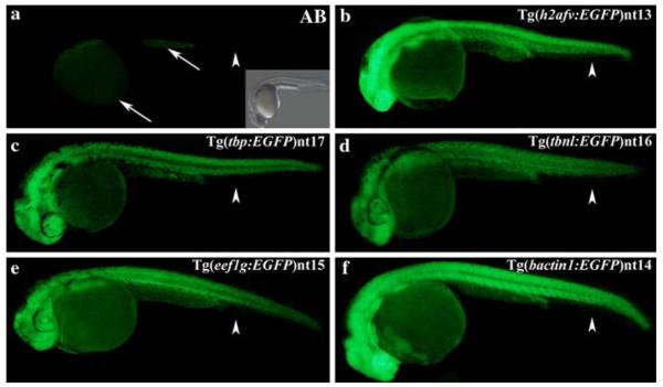
Fig. 3.
EGFP expression between 24 and 36 hpf in the transgenic embryos. The AB embryos possessed only a very low level of autofluorescence in the yolk sac and anal yolk extension at 24–36 hpf (Panel a, arrows). Furthermore, no fluorescence was detected in the fin fold (Panel a, arrowhead). A bright field image showing the AB embryo is inset. The EGFP expression was ubiquitous in the Tg(h2afv:EGFP)nt13, Tg(tbp:EGFP)nt17, Tg(tbnl:EGFP)nt16, Tg(eef1g:EGFP) nt15, and Tg(bactin1:EGFP)nt14 lines (Panels b–f), except for the absence of EGFP expression in the fin folds (arrowheads)

