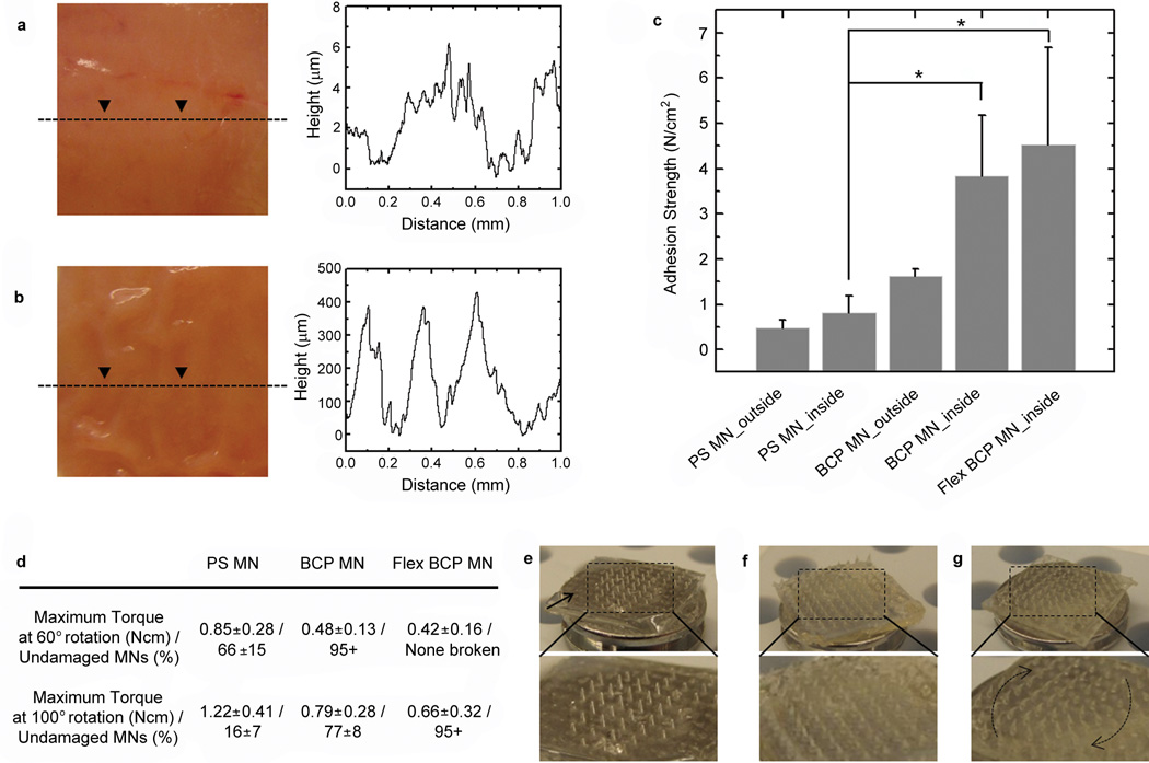
Figure 5. MN adhesive firmly attaches to wet intestine tissue.
a-b, Photographs from a, outer and b, inner (mucosal) surfaces of pig intestine tissue and corresponding depth profile showing topographical roughness. c, Adhesion strength for PS MN (non-swellable) and swellable BCP MN adhesives with a rigid and flexible base following insertion into mucosal and serosal intestine surfaces. The asterisk indicates statistical significance with p < 0.05 (ANOVA with post-hoc Tukey’s HSD test). Error bars represent standard deviation. d, Following penetration into the outer surface of intestine tissue and application of 60° or 100° of rotation, significant damage to PS MN was observed (broken MN), while BCP MN and Flex BCP MN exhibited significantly reduced MN breakage. e-g, Photographs for tilted view of MN arrays after torsion test of 100° rotation using (e) PS MN, (f) BCP MN, and (g) Flex BCP MN adhesives. The tips or entire PS MNs were broken by torsional stress (especially at the edges of the patch marked by arrow), yet BCP MN with rigid and flexible bases showed high resistance to torsional stress by bending in the direction of the shear force.

