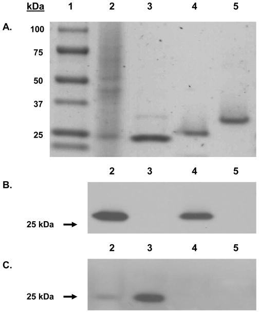
Figure 3. SDS PAGE and western blot analysis of coho salmon OlfGSTs.
(A). Samples were analyzed on a 4–15% gradient SDS-PAGE and proteins visualized by Coomassie Blue staining. Lane 1 contained the protein molecular weight standard; lane 2 contained coho salmon olfactory cytosol (10 μg); lanes 3–5 contained 2.5 μg of recombinant OlfGST pi, rho, and omega respectively. (B–C). Samples were blotted on PVDF membrane and probed with polyclonal GST antisera raised against striped bass GST that recognizes multiple GST forms, including rho of bass [24, 25] and also against a channel catfish GST pi antibody [26]. In both (B) and (C), lane 2 contained coho salmon olfactory cytosol (10 μg); and lanes 3–5 contained 2.5 μg of recombinant OlfGST pi, rho, and omega respectively.

