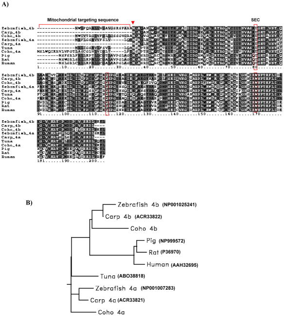
Figure 1.
(A) Predicted amino acid sequence alignment of the two Coho GPx4 isoform with other fish and mammalian GPx4s. The selenocysteine residue is indicated as U in sequence. The three boxes highlight conserved residues at selenocysteine (U), glutamine (Q) and tryptophan (W). Black shading indicates completely conserved residues and grey shading indicates similar residues (similarity threshhold fraction ≥0.7). The arrow indicates the second translational initiation site. Mitochondrial targeting sequences are underlined. (B) Phylogenetic rooted-tree showing the relationship of Coho GPx4 with other fish and mammalian GPx4s. GenBank accession numbers are shown in parentheses. Graph generated from San Diego Biology Workbench 3.2.

