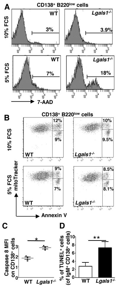
Figure 5. In vitro and in vivo generated Lgals1−/− plasmablasts are more susceptible to apoptosis than WT plasmablasts.
(A-C) B220+ splenic B cells from WT and Lgals1−/− mice were cultured with 1μg/ml LPS during 3 days in complete medium containing 10% or 5% FCS. (A) Quantification of apoptotic B220lowCD138+ plasmablasts was determined by 7-AAD incorporation. (B) Quantification of mitochondrial depolarization and phosphatidyl serine externalization on B220lowCD138+ plasmablasts using mitotracker and annexin V staining. Lived cells are mitotrackerhigh/annexinV−, cells with externalized phosphatidyl serines are mitotrackerhigh/annexinV+ and cells with a decreased mitochondirial potential are mitotrackerlow/annexinV+. (C) Quantification of caspase 8 activation. Fold changes in active caspase 8 MFI in B220lowCD138+ plasmablasts generated in culture medium supplemented with 10% or 5% FCS are represented. Data in A and B are representative of 3 independent experiments. (D) WT and Lgals1−/− mice were immunized with NP-KLH in alum (T-dependent Ag), 6 mice for each group, and the percentage of splenic apoptotic cells within IgM+CD138+ cells was quantified using TUNEL assays at day 21. For WT and Lgas1−/− mice 3893 and 2154 cells were counted, respectively.
* and ** indicate significance versus WT mice with p<0.05 and <0.005 respectively.

