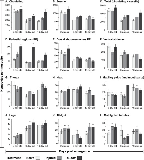
Figure 1.
Systemic hemocyte numbers decrease with age but increase after infection. Number of circulating (A), sessile (B) and total (C; circulating and sessile) hemocytes at different ages in naïve mosquitoes (light gray), and mosquitoes that had been either injured (medium gray) or infected with E. coli (dark gray) for 24 hours. (D) Number of sessile hemocytes attached to the dorsal abdominal wall at the location of the ostia (periostial regions). (E) Number of sessile hemocytes attached to the dorsal abdominal wall minus the periostial hemocytes. (F) Number of sessile hemocytes attached to the ventral abdominal wall. (G-L) Number of sessile hemocytes in the thorax (G), head (H), maxillary palps (I), legs (J), midgut (K), and Malpighian tubules (L). Column heights mark the average number of hemocytes per mosquito, and whiskers denote the standard error of the mean.

