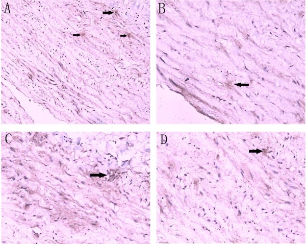
Figure 3.
Immunohistochemical staining. Ascending aorta specimen in the entry site of the dissection from a AD patients were sectioned and labeled with MMP-12 antibodies as indicated. Sections were developed with alkaline phosphatase anti-alkaline phosphatase (APAAP) techniques and counterstained with hematoxylin. Total cells and positive cells were counted under the microscope. Arrows indicate examples of MMP-12 positive cells, A (magnification×200), B, C, D (magnification×400).

