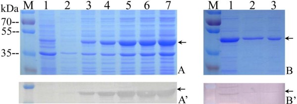
Figure 1.

Identification and purification of recombinant VP6 protein in vitro. A: SDS-PAGE analysis of induced recombinant VP6 expression. M, standard protein marker; Lane 1, expression of pREST empty vector induced by IPTG for 3 h; 2–7, expressed recombinant VP6 cell lysate pellet induced by IPTG for 0, 1, 2, 3, 4, 5 h respectively. A’: Western blotting analysis corresponding to lane 1–7 in A with His-tag monoclonal antibody. B: SDS-PAGE analysis of the purified rVP6 protein. Lane 1, recombinant VP6 cell lysate; Lane 2, 3, purified rVP6. B’: Western blot analysis of purified rVP6 protein matching lane 1–3 in B with rabbit anti-GCRV antibody. Arrows indicate rVP6 protein.
