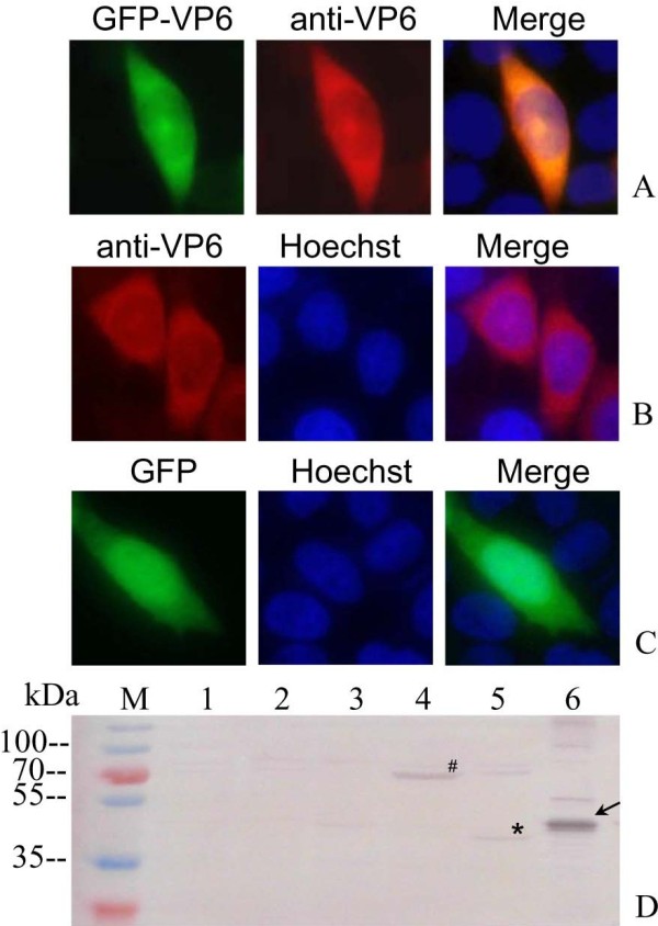
Figure 3.

Indirect IF and IB assays of the VP6 protein in transfected cells. A, B and C: Intracellular localization of single VP6 protein in transfected cells, Vero cells were transfected with plasmid pEGFP-VP6 (A), pCI-VP6 (B) and pEGFP (C) respectively for 24 h, and then immunostained with mouse anti-VP6 polyclonal serum followed by Alexa Fluor® 568 donkey anti-mouse IgG (red) for EGFP-VP6 and VP6. The nuclei were visualized by counterstaining with Hoechst. D: IB analysis of expression of pEGFP-VP6 and pCI-VP6 in transfected Vero cells. M, standard protein marker; Lane1, mock transfected Vero cell lysates; 2–5, transfected Vero cell lysates with pCI-neo empty vector, pEGFP-C1 empty vector, pEGFP-VP6 (71 kDa) plasmid, pCI-VP6 (45 kDa) plasmid respectively; Lane 6, recombinant His-tag fusion VP6 protein (48 kDa) as positive control. #: GFP-VP6, *: VP6, Arrow presents rVP6 protein.
