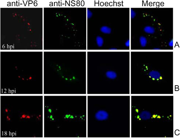
Figure 4.

Subcellular colocalization of the VP6 and NS80 proteins in virus infected cells. IF microscopy of GCRV infected CIK cells at 6 h (A), 12 h (B), 18 h (C), p.i. The subcellular localizations of VP6 and NS80 were detected by immunostaining with mouse anti-VP6 and rabbit anti-NS80 polyclonal antibodies followed by Alexa Fluor® 568 donkey anti-mouse IgG (red) and Alexa Fluor® 488 donkey anti-rabbit IgG (Green). Nuclei were counterstained with Hoechst (blue).
