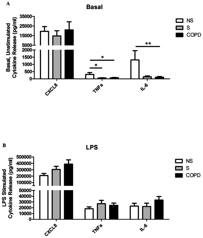
Figure 1. Baseline and LPS-stimulated characteristics of alveolar macrophage supernatants.
Lung macrophages were incubated either with or without LPS (1 µg/ml) for 24 h with 17-BMP (0.01–100 nM). Data shown are mean ± SEM for CXCL8, TNFα and IL-6 ELISAs of baseline (A) and LPS-stimulated (B) supernatants. COPD (n = 25), S (n = 11) and NS (n = 8) shown. One-way ANOVAs were performed on all data sets, and when P<0.05 two-tailed t-tests were subsequently performed which are shown on the graph; * P<0.05, ** P<0.01.

