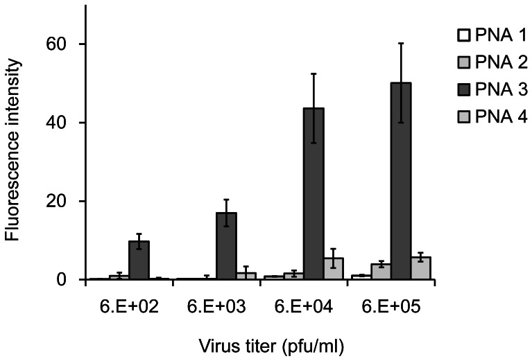
Figure 3. Fluorescence detection of the influenza A/Osaka/180/2009(H1N1) virus genome on PNA 1–4-immobilized plates.
Conditions: 0.5 µg of PNA was immobilized on each well. Virus incubation: 100 µl of indicated virus solution, 1 h, r.t. Antibodies: 0.1 µg mouse monoclonal influenza A virus nucleoprotein primary antibody and goat anti-mouse IgG secondary antibody conjugated with horseradish peroxidase in each well, 2 h, r.t. Incubation of 3,3′,5,5′-tetramethyl-benzidene with horseradish peroxidase: 10 min, r.t. Fluorescence detection: excitation 430 nm, emission 503 nm.

