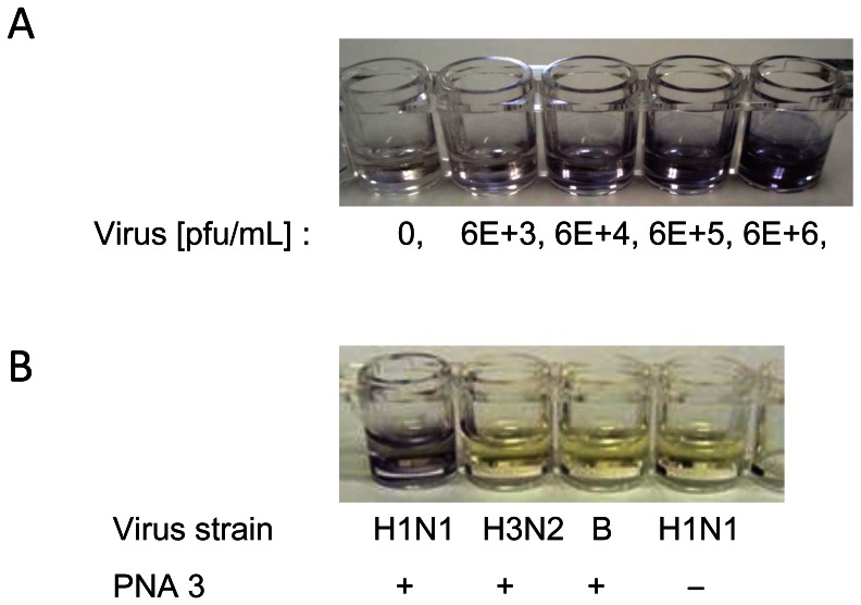
Figure 5. Visual detection of the NS gene of influenza A/Osaka/180/2009 (H1N1pdm) virus in wells containing immobilized PNA 3.
A) Conditions: as for Fig. 3, but the virus concentration was 10-fold higher and the secondary antibody and the substrate were changed to anti-mouse IgG conjugated with alkaline phosphatase, and 5-bromo-4-chloro-3′-indolyphosphate and nitro-blue tetrazolium (BCIP/NBT), respectively. Incubation of BCIP/NBT with ALP: 60 min at r.t. B) Conditions: as for Fig. 4, except that the secondary antibody and the substrate were changed to anti-mouse IgG conjugated with alkaline phosphatase, and 5-bromo-4-chloro-3′-indolyphosphate and nitro-blue tetrazolium (BCIP/NBT), respectively. Incubation of BCIP/NBT with ALP: 60 min at r.t.

