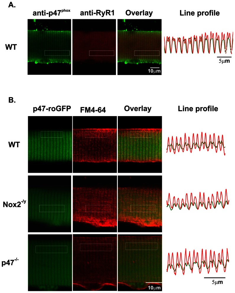
Figure 5. Localization of p47-roGFP in skeletal muscle.
(A) Immunostaining of enzymatically dissociated single FDB myofibers for endogenous p47phox and the ryanodine receptor shows that p47phox co-localizes with the ryanodine receptor at the triad. (B) Fluorescent live cell image of an FDB electroporated with p47-roGFP and counter stained with the membrane and t-tubule dye FM4-64® shows that p47-roGFP is localized at the t-tubule. The line plots represent the longitudinal spatial profile of fluorescence averaged over the transverse direction within the boxed regions.

