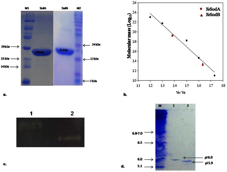
Figure 3. Molecular weight, activity and pI analysis of recombinant SODs:
(a) SDS–PAGE of recombinant YeSodA and YeSodB expressed in pET 28a (+) (samples were resolved on 15% polyacrylamide gel and stained with Coomassie Brilliant Blue R-250). The purified SodA and SodB showed a single band each of 23 KDa and 21 kDa respectively. M1 and M2: Protein marker; Lane 1: SodA; Lane 2 SodB. (b) Molecular weight determination of YeSodA (82 kDa) and YeSodB (21 kDa) by Sephacryl S-200 molecular sieve chromatography. The molecular weight of marker proteins (SigmaAldrich) were as follows: β-Amylase (200 kDa), Alcohol dehydrogenase (150 kDa), BSA (66 kDa), Carbonic anhydrase (29 kDa) and Cytochrome C (12.4 kDa). (c) Zymogram analysis showing achromatic bands of YeSodA and YeSodB against a dark background. Lane 1: YeSodA; Lane 2: YeSodB. (d) Isoelectric point (pI) of purified recombinant YeSodA and YeSodB stained with coomassie brilliant blue. M: pI marker; Lane 1: YeSodA; Lane 2: YeSodB.

