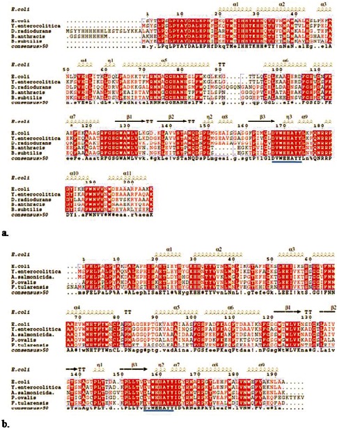
Figure 5. Sequence homology:

Multiple sequence alignment (MSA) of (a) YeSodA with E. coli (PDB id: 1VEW), Deinococcus radiodurans (PDB id: 2CDY), B. anthracis (PDB id: 1XUQ) and B. subtilis (PDB id: 2RCV); (b) YeSodB with E. coli (PDB id: 2NYB), Aliivibrio salmonicida (PDB id: 2W7W), Pseudomonas ovalis (PDB id: 1DT0) and Francisella tularensis (PDB id: 3H1S) drawn using ESPript 2.2. Symbols α and β indicate alpha helices and beta sheets, respectively; η represents turns and TT denotes sharp turns in the structure.
