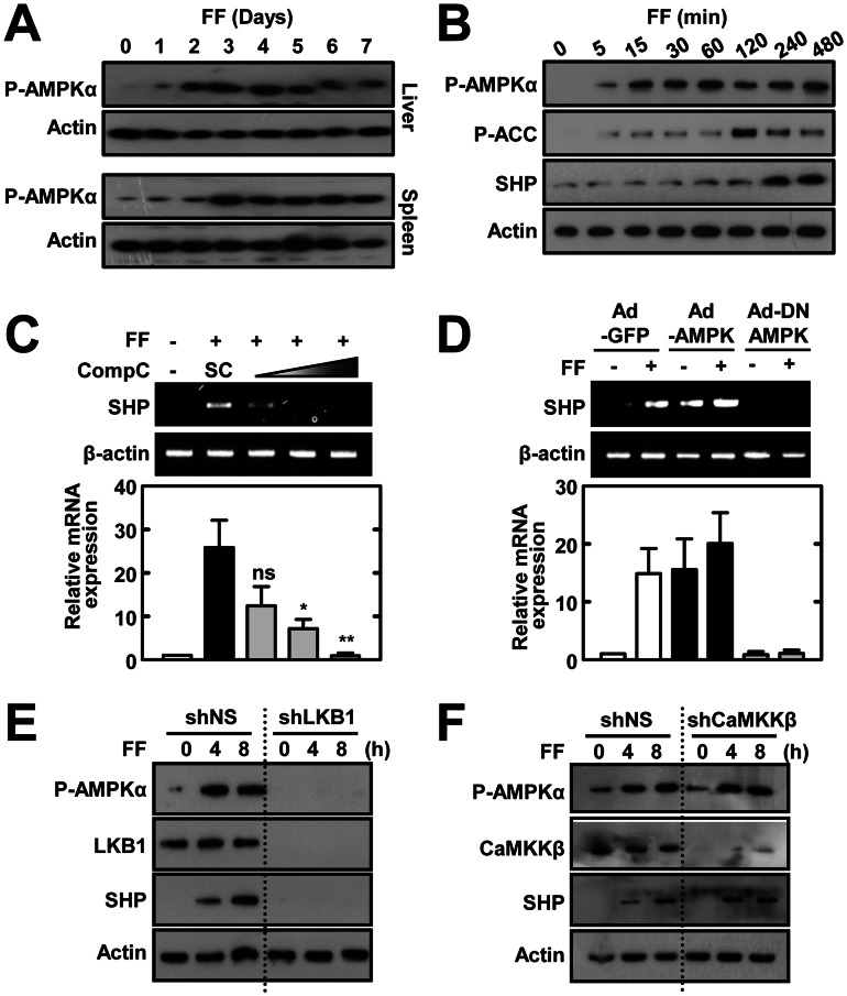
Figure 5. Fenofibrate induces SHP expression through LKB1-dependent AMPK signaling.
(A) WT mice (n = 3) received fenofibrate (100 mg/kg; administrated orally) or vehicle only for the indicated period. The expression of phosphorylated AMPKα in liver and spleen was assessed by immunoblotting (IB). Whole cell lysates (WCL) were used for IB with anti-Actin. (B) BMDMs were treated with fenofibrate (50 µM) for various times, followed by IB with phosphorylated forms of AMPKα, ACC, and SHP. WCL were used for IB with anti-β-actin as the loading control. (C) BMDMs were treated with fenofibrate (50 µM for 6 h) in the presence or absence of the AMPK inhibitor compound C (Comp C; 5, 10, or 25 µM). Total RNA was extracted from WCL and used for RT-PCR analyses of Shp and Actb mRNA; below, densitometry. (D) BMDMs were transduced for 48 h with adenovirus encoding GFP only (Ad-GFP), constitutively active AMPK (Ad-AMPK), or dominant negative AMPK (Ad-DN-AMPK; MOI = 10) and were then treated with fenofibrate (50 µM) for 4 h. Total RNA was extracted from WCL and analyzed by semi-quantitative RT-PCR for Shp and Actb mRNA. Representative gel images (top); densitometric analyses (bottom). (E and F) BMDMs were transduced with shNS or shLKB1 (E) or shCAMKKβ (F) prior to treatment with fenofibrate (50 µM) for the indicated time periods, followed by IB with antibodies against the phosphorylated forms of AMPKα and SHP. WCL were used for IB with anti-β-actin as the loading control. Total LKB1 (D) and CAMKKβ (E) protein expression was measured for transduction efficiency of lentiviral vectors. The data are representative of at least three independent experiments with similar results. Quantitative data are shown as the mean ± SD of three experiments (C and D bottom). Statistical differences (*, p<0.05; **, p<0.01), as compared to the control cultures, are indicated (paired t-test with Bonferroni adjustment). FF, fenofibrate. n.s., non-specific.

