
Figure 2. MARCKS participates in acrosomal exocytosis.
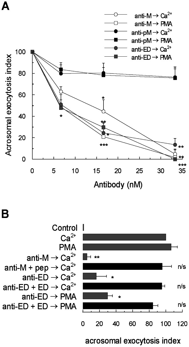
(A) Permeabilized sperm were incubated for 15 minutes at 37°C in the presence of increasing concentrations of anti-MARCKS N-19 antibody (anti-M, white symbols), anti-MARCKS ED (anti-ED, gray symbols) and anti-phospho-MARCKS (anti-pM, black symbols). Acrosomal exocytosis was initiated by adding 10 µM free Ca2+ (circles) or 200 nM PMA (squares). For all conditions, the incubation continued for an additional 15 minutes and acrosomes were evaluated by lectin binding. (B) Permeabilized sperm were incubated for 15 minutes at 37°C in the presence of 33 nM anti-MARCKS N-19 antibody (anti-M) preincubated with an excess (1∶ 10) of blocking peptide MARCKS N-19 (anti-M+pep) and in the presence of 33 nM anti-MARCKS ED antibody (anti-ED) preincubated with an excess (1∶10) of MARCKS effector domain (anti-ED+ED). Acrosomal exocytosis was then initiated by adding 10 µM free Ca2+ or 200 nM PMA and the incubation continued for an additional 15 minutes (black bar). Control experimental conditions (gray bars) include background acrosomal exocytosis in the absence of any stimulation (control), acrosomal exocytosis stimulated by 10 µM free Ca2+ (Ca2+) and by 200 nM PMA (PMA) and the effect of antibodies without peptide preincubation anti-MARCKS N-19 (anti-M) and anti-MARCKS ED (anti-ED). The percentage of reacted sperm was normalized as described in Materials and Methods. The data represent the means±SEM of at least three independent experiments. The asterisks indicate significant differences from similar conditions stimulated with Ca2+ (**, p<0.01; *, p< 0.05); n/s, not significant difference.