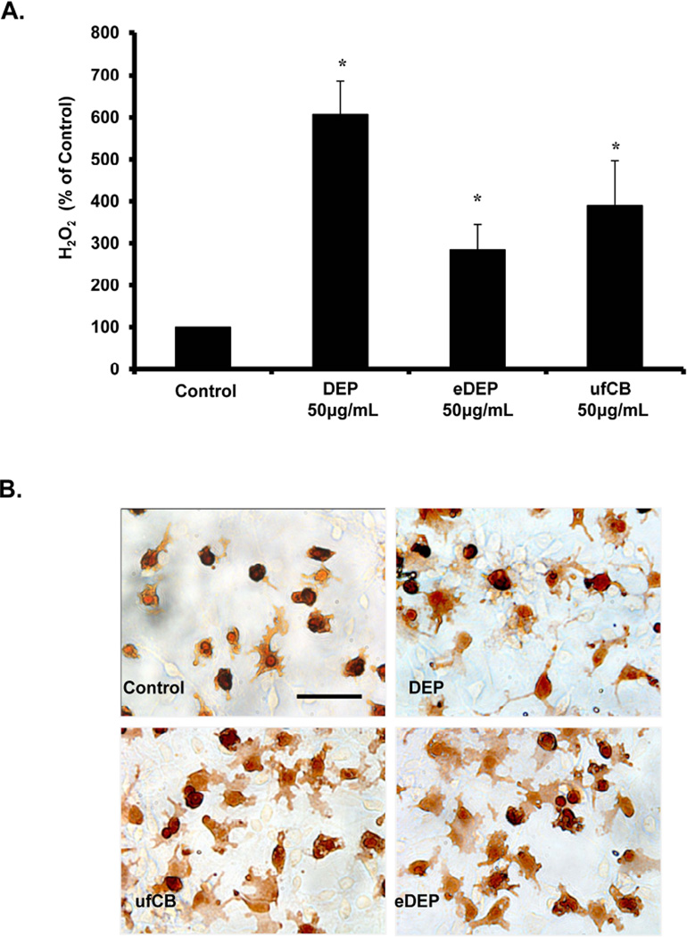
Figure 1. Components of diesel exhaust particles (DEP) activate microglia.
(A) HAPI microglia cells were treated with DEP (50µg/ml), DEP extract (eDEP, from 50µg/ml DEP), and carbon black (ufCB, 50 µg/ml). The production of hydrogen peroxide (H2O2) was measured by the catalase-inhibitable fluorescence. Samples were run in triplicates and the data are the result of 4 independent experiments (n=4). Results are expressed as percent of control and represent the mean ± SEM. The raw data (fluorescence) for the control treatment range from 580 – 906 across experimental replicates. An asterisks indicates a significant difference from control (1 Way ANOVA, p<0.05). (B) Primary neuron-glia cultures were treated with DEP (50µg/ml), eDEP (from 50µg/ml DEP), and ufCB (50 µg/ml) for 9 hr and stained with the IBA-1 antibody. Microglial activation in response to the DEP components is depicted by an increase in number of stained cells, enlarged size of stained cells, and irregular amoeboid morphology. Representative images from the culture are shown from three independent experiments (n=3). Images were taken at 400× and the scale bar depicts 20µM.

