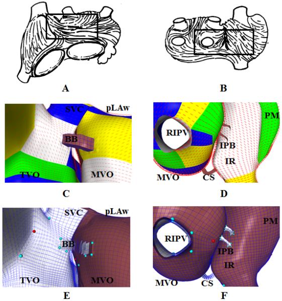
Figure 6.
Fiber orientations in a tricubic Hermite model. Images (A) and (B) are depictions of typical fiber orientations from explanted human atria, reprinted from Wang, Br. Heart J., 1995 with permission. The regions indicated by boxes are enlarged in (C) and (D), in which a qualitatively-matching fiber pattern is displayed. Each block of color indicates a region with consistent coordinate axes (a single topological region). See Results, Section 3.3 for details. Images (E) and (F) are equivalent views of the tricubic Hermite hexahedral model. The epicardial surface is colored brown and the endocardial surface is colored white. Valence 3 vertices are colored red, and valence 5 vertices are colored teal. SVC = Superior Vena Cava; BB = Bachmann's bundle; TVO = Tricuspid valve orifice; pLAw = Posterior left atrial wall; MVO = Mitral valve orifice; RIPV = Right inferior pulmonary vein; CS = Coronary sinus; IR = Intercaval region; PM = Pectinate muscles, IPB = Inferoposterior bridge.

