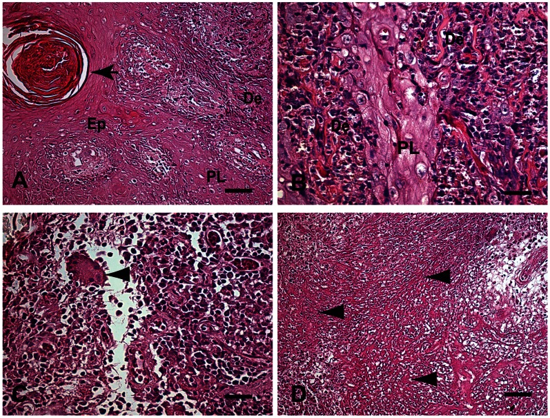
Figure 1. A–D: Fragment of skin of a patient with LCL Caratinga, MG, Brazil.
(A) Changes observed in the epidermis were intense acanthosis (AC) and papillomatosis (PL). Pearl corneas can also be seen (black arrow). Finger-like projections of epidermis into the dermis layer, papillomatosis (PL) Bar = 32 µm, (B) Higher magnification shows thickening of the spinous (acanthosis) layer due to proliferation of epidermal cells (arrowhead) leading to papillomatosis (PL). Bar = 16 µm, (C) Higher magnification showing the inflammatory infiltrate of mononuclear cells (plasma cells, macrophages and lymphocytes) in the dermis. Note Langhans-type giant cell formation, but without a typical granuloma formation (arrowhead). Bar = 16 µm. (D) Eosinophilic necrotic area in the dermis with fragmented collagen fibers resembling fibrinoid necrosis (arrowheads) Bar = 64 µm. Hematoxylin-eosin staining. Epithelium (Ep), Dermis (De), Papillomatosis (PL).

