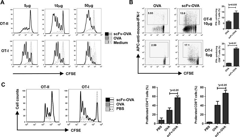
Figure 3. T-cell responses mediated by scFv-OVA.
(A, B) Splenocytes from OT-I or OT-II mice were labeled with CFSE and then stimulated with different concentrations of OVA or scFv-OVA for 3 days. The turnover of T cells and intracellular IFN-γ staining from splenic cells were examined by flow cytometer. Cells were gated on CD8+ or CD4+ populations. T-cell proliferation shown in overlay histogram was from medium, OVA or scFv-OVA (A). The IFN-γ production of CD4+and CD8+ was shown in (B). (C) 2×10⁶ CFSE-labeled naive CD4 OT-II or CD8 OT-I T cells were i.v. adoptively transferred into naive C57Bl/6 mice (n=3). The next day, mice were injected with a single dose of OVA or scFv-OVA (for OT- I T cells, 5 μg protein/mouse; for OT- II T cells, 20 μg protein/mouse) or PBS. Recipient mice were killed after 3 days and turnover of T cells from splenic cells was examined by flow cytometer. Cells were gated on CFSE-positive population. T-cell proliferation shown in overlay histogram was from PBS, OVA, or scFv-OVA. Representative of 3 experiments.

