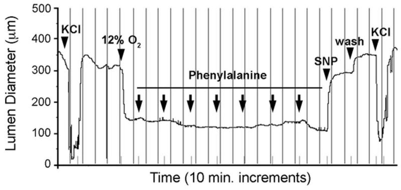
Figure 3.

Response of the isolated murine ductus arteriosus to phenylalanine. Representative tracing of a term gestation mouse ductus that was mounted in a pressure myography chamber under deoxygenated conditions. Submaximal constriction of the ductus lumen diameter by oxygen was not overcome by exposure to increasing concentrations of phenylalanine (10−9 M to 10−3 M). In contrast, exposure to the NO donor SNP (10−5 M) produced marked dilation and return to resting baseline dimensions. Terminal exposure to 50mM KCl confirmed responsiveness of the preparation at completion of the study. Vertical lines represent 10 minute intervals. Arrowheads indicate serial 10-fold increases in phenylalanine concentration in the perfusion bath.
