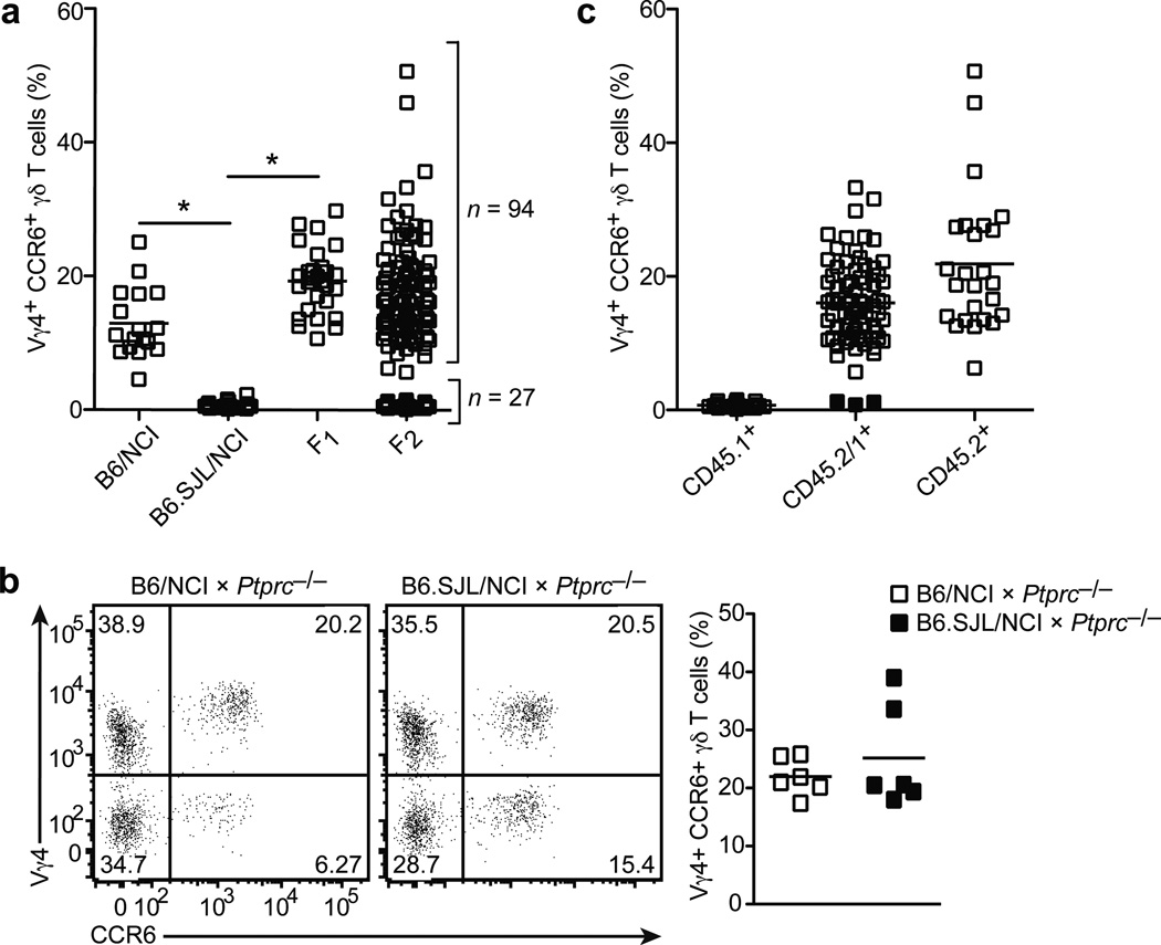
Figure 2. Vγ4+ γδT17 cell deficiency is an autosomal, recessive trait controlled by a single locus.
(a) Quantification of LN Vγ4+CCR6+ γδ T cell frequency (plotted as % of total γδ T cells) in mice of the indicated type. (b) Flow cytometric detection of Vγ4+CCR6+ γδ T cells in digested lymph node cell suspensions from B6/NCI and B6.SJL/NCI × Ptprc−/− mice (3 to 6 weeks of age), gated on total γδ T cells (left panel); quantification of LN Vγ4+CCR6+ γδ T cell frequency (plotted as % of total γδ T cells) in mice of the indicated type (right panel). (c) Quantification of LN Vγ4+CCR6+ γδ T cell frequency (plotted as % of total γδ T cells) in F2 mice that are CD45.1+, CD45.1+/2+, and CD45.2+. The three recombination events are indicated by the filled squares. Each symbol represents an individual mouse; horizontal bars represent the mean (a–c). *P≤0.0001. Data are representative of 18 experiments with at least 15 mice of each type (a,c) and two experiments with 6 mice (b).

