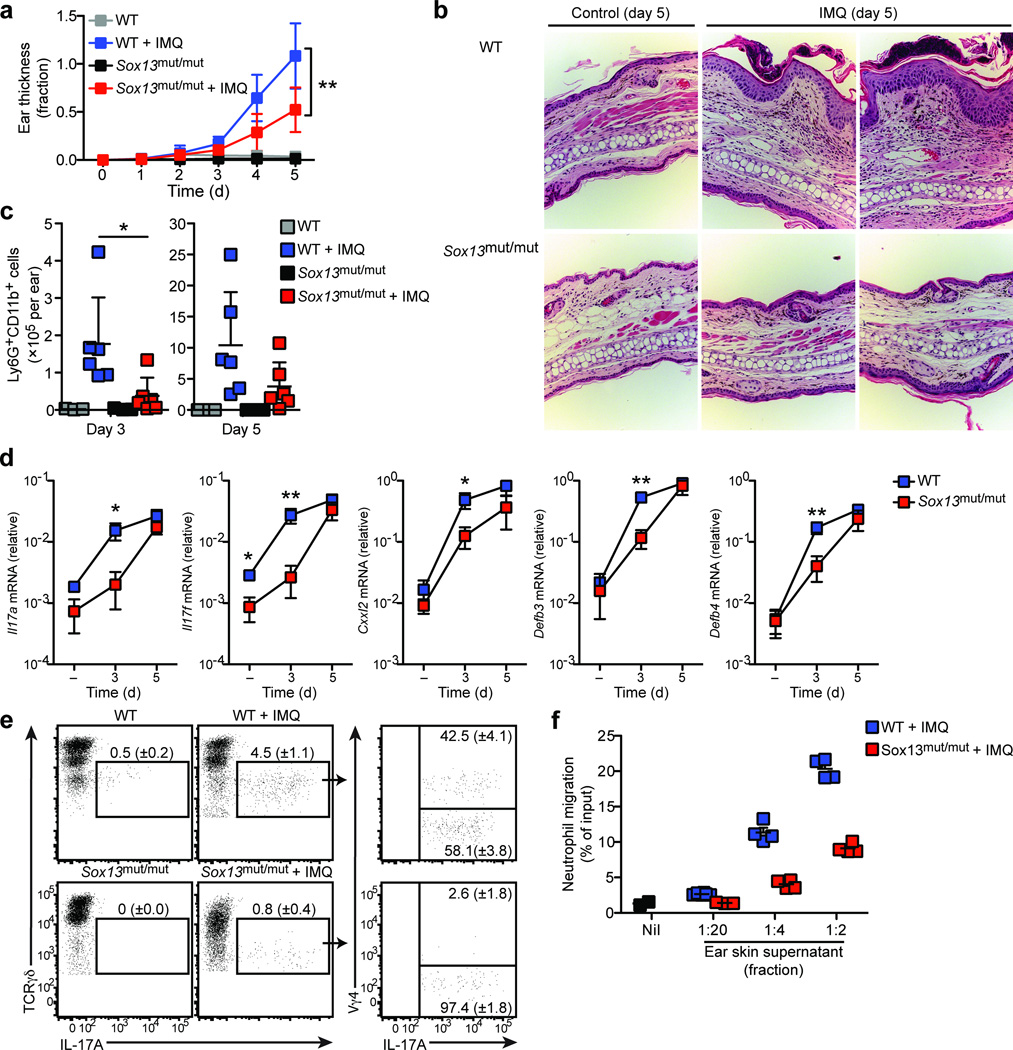
Figure 6. Sox13mut/mut (B6.SJL/NCI) mice are protected from psoriasis-like dermatitis.
(a) Quantification of ear skin thickness, plotted as fraction increase relative to baseline (day 0), of WT (B6/NCI) and Sox13mut/mut (B6.SJL/NCI) mice treated with imiquimod or control cream daily for 5 days. Boxes represent the mean (± s.d.). (b) H&E staining of ear skin from WT and Sox13mut/mut mice treated per (a) for 5 days. (c) Quantification of Ly6G+CD11b+ neutrophils in ear skin cell suspensions from WT and Sox13mut/mut mice treated per (a) for 3 or 5 days. Each symbol represents an individual mouse; horizontal bars represent the mean (± s.d.). (d) RT-PCR quantification of ear skin mRNA from WT and Sox13mut/mut mice treated with control (−) or imiquimod cream for 3 or 5 days. Boxes represent the mean (± s.d.). (e) Intracellular IL-17A staining of ear skin cell suspensions from WT and Sox13mut/mut mice treated as in (a) for 3 days and digested in the presence of Brefeldin A, gated on total γδ T cells. Mean (± s.d.) is indicated. (f) Transwell assay of neutrophil migration to ear skin supernatants prepared from WT and Sox13mut/mut mice treated as in (a) for 3 days. Each symbol represents migration from an individual transwell, horizontal bars represent the mean (± s.d.). *P≤0.05, **P≤0.01. Data are representative of three experiments with 3–6 mice (a–d), two experiments with 2–5 mice (e), and four experiments with 9 mice (f).

