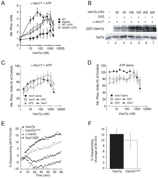
Figure 1. Non-canonical SNARE complexes stall at a hemifusion intermediate.

(A) Individual fusion reactions were blocked at the priming stage by treating vacuoles with 85 μg/ml anti-Sec17p antibody for 15 minutes. Recombinant Vam7p (closed circles) or Vam7pQ283R (closed squares) was added to bypass the priming block. In parallel, Vam7p and Vam7pQ283R were added to reactions blocked with anti-Sec17p antibody and before treatment with CPZ. (B) Fusion reactions were treated with anti-Sec17p antibody in the presence or absence of CPZ. Next, recombinant Vam7p was added to reactions at the indicated concentrations. After incubation (10 minutes, 27°C), membranes were pelleted (16,000 g, 10 minutes, 4°C) and the unbound Vam7p was discarded with the supernatant. Bound Vam7p was examined by immunoblotting. Membranes were also probed for Ypt7p as a control for loading. (C) Fusion reactions were treated with anti-Sec17 antibody after which Vam7p alone (closed circles) was added to bypass the fusion block. In parallel, reactions were treated with CPZ either before (open triangles) or after addition of Vam7p (open squares). (D) Uninhibited fusion reactions were treated with Vam7p alone (closed circles). In parallel, reactions were treated with CPZ either before (open triangles) or after addition of Vam7p (open squares). (E) Lipid mixing (hemifusion) assays were performed using vacuoles blocked with anti-Sec17p at 4°C. After 15 minutes, 400 nM Vam7p (WT or Q283R) was added to the indicated reactions while on ice prior to transferring to the plate to 27°C and further incubation (60 minutes). The increase in fluorescence occurred when the outer leaflets of vacuoles were mixed during hemifusion. Shown is a representative of 3 trials. (F) Shown is the average lipid dequenching at 60 minutes. Error bars indicate SEM (n=3).
