
Figure 1. Computation of the DRF.
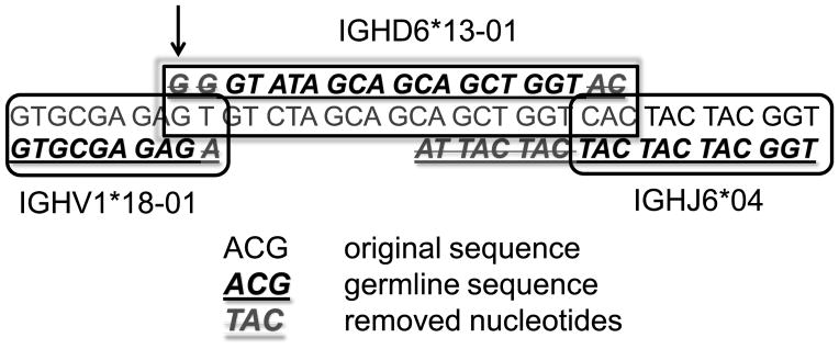
We first compute the position of VH (from ACGA to GAGA in the first row and the beginning of the second row). The upper line is the full sequence, and the lower line is the germline gene (VH. in this case). We then compute how the codons are aligned with respect to the beginning of VH (see for example the GAG at the end of V). On the 3' end of the sequence we compute the position of JH (lower right hand part of figure). Again the sequence is above the germline JH gene. In between, we compute DH (central lower part). The sequence above the text is the germline and we compute its position versus the reading frame of VH. In this case it starts at the third RF (the first G of the germline DH, indicated by the arrow, is above a G in the third reading frame of the VH germline). Crossed out nucleotides are germline nucleotides that were removed from either VH, DH or JH in the join region.